|
|
|
|
|
| Health
Check-up Plan | Individual Diagnostic Test | FAQs| |
|
|
| |
2D Echo
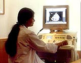
Two-dimensional echocardiography
can provide excellent images of the heart, Para cardiac
structures, and the great vessels. During a standard
echo, the sound waves are directed to the heart from
a small hand-held device called a transducer, which
sends and receives signals. Heart walls and valves
reflect part of the sound waves back to the transducer
to produce pictures of the heart. These images appear
in black and white and in color on a TV screen. They're
selectively recorded on videotape and special paper,
and later reviewed and interpreted by a cardiologist
(heart specialist).
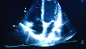  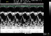 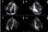
From the pictures it is possible
to measure the size of each part of your heart, to
study motion and appearance of the valves and the
function of the heart muscle. Your physician uses
the measurements to determine how your heart is working
and whether or not any abnormalities are present.
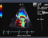
A Doppler echo is often done at
the same time in order to determine how the blood
flows in your heart. The swishing sounds you hear
during the test indicate blood flowing through the
valves and chambers.
Highlights:
- Carotid Colour Doppler
- Peripheral arterial & venous colour Doppler
- Abdominal Colour Doppler
- Pregnancy Colour Doppler
Patient Benefits:
- Faster Examination
- High resolution images for detection of subtle
abnormalities.
- Vascular information
|
|
|
|
| Dental
Care |
Cosmetic
Surgeries |
Health
Check up Plan |
|
|
|