|
|
|
|
|
| Health
Check-up Plan | Individual Diagnostic Test | FAQs| |
|
|
| |
ECG
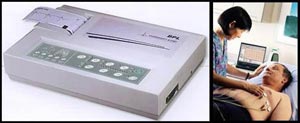 Electrocardiography
(ECG or EKG) is the recording
of the electrical activity of the heart over time
via skin electrodes.[1] It is a noninvasive recording
produced by an electrocardiograph. The etymology of
the word is derived from electro, because it is related
to electrical activity, cardio, Greek for heart, graph,
a Greek root meaning "to write"
Electrocardiography
(ECG or EKG) is the recording
of the electrical activity of the heart over time
via skin electrodes.[1] It is a noninvasive recording
produced by an electrocardiograph. The etymology of
the word is derived from electro, because it is related
to electrical activity, cardio, Greek for heart, graph,
a Greek root meaning "to write"
Electrical impulses in the heart
originate in the sinoatrial node and travel through
conducting system to the heart muscle. The impulses
stimulate the muscle fibers to contract and thus producing
the systole. The electrical waves can be measured
at selectively placed electrodes (electrical contacts)
on the skin. Electrodes on different sides of the
heart measure the activity of different parts of the
heart muscle. An ECG displays the voltage between
pairs of these electrodes, and the muscle activity
that they measure, from different directions, also
understood as vectors. This display indicates the
overall rhythm of the heart and weaknesses in different
parts of the heart muscle.
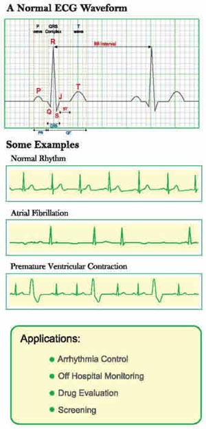
Helps In Diagnosis of:
- It is the best way to measure
and diagnose abnormal rhythms of the heart,
- Particularly abnormal rhythms
caused by damage to the conductive tissue that carries
electrical signals, or abnormal rhythms caused by
levels of dissolved salts (electrolytes), such as
potassium, that are too high or low.
- In myocardial infarction (MI),
the ECG can identify damaged heart muscle. But it
can only identify damage to muscle in certain areas,
so it can't rule out damage in other areas.
- The ECG cannot reliably measure
the pumping ability of the heart; for which ultrasound-based
(echocardiography) or nuclear medicine tests are
used.
Procedure:
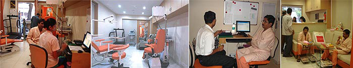
You will be brought to the ECG department
in the centre. The technician will ask you to take
off your clothing from the waist up, put on a gown,
and lie down on a small bed. The technician will place
a small electrode (a small self-sticking plastic patch)
on each of your arms and legs and across six areas
on your chest. You need to lie still for the minute
or two that it takes the machine to make a record.
|
|
|
|
| Dental
Care |
Cosmetic
Surgeries |
Health
Check up Plan |
|
|
|